
Large scale introduction of CT lung screening will put an enormous burden on radiologists. The ever increasing number of patients will lead to more cases that require reading. Despite these added efforts, lung screening programs will need to maintain a quality environment.
Veolity™ is the reading platform for high-throughput environments. Built upon years of clinical and technical experience from large screening trials, it provides important innovations to improve the reading workflow and diagnostic quality.
The key benefits of Veolity are:
Highly optimized reading workflow
Veolity organizes the reading workflow allowing to quickly generate a report. From preparing optimized worklists to fast opening times achieved by automatic preloading, everything is prepared to immediately start reading. Additionally, all images are sorted into an intelligent layout. The visualization adapts to the situation, e.g. automatically switching on a slab mode when searching for nodules, while zooming into segmented structures allowing a detailed analysis.
Overall, Veolity provides the next generation reading workflow.
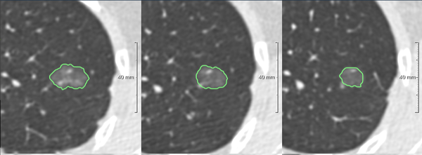
One-click nodule segmentation
Sophisticated one-click segmentation provides automated measurements such as diameter and 3D volume. The default segmentation delivers accurate results within an instant and was validated on thousands of cases. If the user needs further adjustments on the automatic results, easy tools allow changing the segmentation quickly.
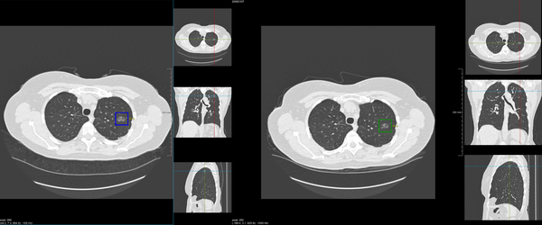
Seamless integration of prior cases
When sending a new case to Veolity, it automatically checks for any existing priors on PACS. All cases of a patient are run through the accurate registration algorithms, generating a complete anatomical mapping. This allows not only to work with the images synchronously, but also to propagate any prior finding to the current case.
Fully integrated solid pulmonary nodule CAD
Veolity is designed to maximize efficiency by assisting in cumbersome tasks such as the identification of suspicious regions. Each case is processed by an FDA approved CAD algorithm detecting actionable lung nodules and highlighted to the user. Extensive validation of the algorithm showed a high sensitivity with a low false positive rate, making it the second pair of eyes for the radiologist.
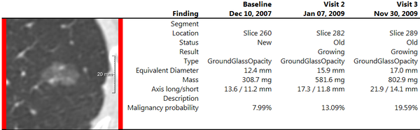
State-of-the-art-reporting
Every reading workflow needs to result in a perfectly structured report that determines patient follow-up.
The Veolity report is based on the LungRADS criteria and includes all reported nodules over time. Comprehensive result tables allow to get a quick overview about the patient. Additionally, current guidelines such as nodule malignancy prediction1, emphysema and calcium scoring complete the overall picture.
1A. McWilliams, M.C. Tammemagi, J.R. Mayo et al. Probability of cancer in pulmonary nodules detected on first screening CT N Engl J Med, 369 (10) (2013), pp. 910–919
Clinical integration
Veolity works with current CT scanners from all major vendors. It offers a variety of deployment options and scaling possibilities so that it can be set-up to match every clinical environment. From single workstation to a high-throughput client-server configuration, Veolity integrates seamlessly into the existing workflow.
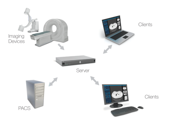
For more information on how to integrate Veolity in your environment, please contact us via sales(at)veolity.com or use the contact form.